


The ankle joint is often injured due to heavy loads. Such a diagnosis of ankle joint disease is not uncommon. It can be placed regardless of the age and gender of the patient. What is ankle joint disease and how is it treated?
what is this?
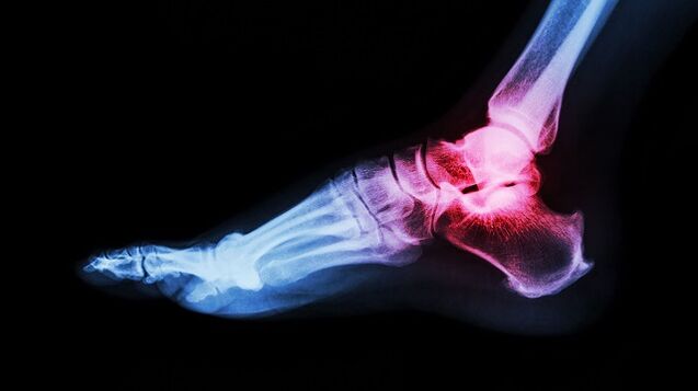
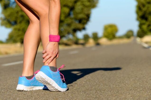
The ankle is under a huge load. Its function is to keep the body upright. Thanks to him, walking and running alone. Due to the violation of the ankle system, it is extremely difficult to live a familiar lifestyle. What is interfering with the work of the ankle?
Ankle joint disease, what is going on? This is a chronic joint disease characterized by degenerative disease. In articular cartilage, irreversible processes are triggered, which can lead to serious complications.
Ankle joint disease gradually develops. A healthy joint surface is elastic and smooth. They provide cushioning under heavy loads and glide smoothly while driving. With pathology, tissue nutrition and metabolism are disturbed. The surface of the joint becomes inelastic and rough. During exercise, cartilage touches each other, causing inflammation. When lifting weights, the main load falls on the bones, which can lead to degenerative diseases.
Lack of treatment can lead to more serious diseases. In 3-4 stages, cartilage and tissue damage is observed. Inflammation of the synovium. The joints become unstable. Violation of support function. All these violations add up to make it impossible to move.
Arthropathy (osteoarthritis) is one of the most common joint diseases and affects a considerable number of people.
Causes and risk factors
What is ankle joint disease, we have sorted it out. Now let us find out what is the root cause of it. Ankle joint disease is considered a senile disease. This is due to age-related physical changes. Cartilage becomes thinner and bones become unstable and fragile. However, in the past decade, the diagnosis of ankle joint disease has become younger. Such statistics are disappointing because many patients ignore the first signs of disease. Late diagnosis always threatens the development of serious complications.
Precipitating factors include:
- dislocation;
- Bruise
- Inflammatory diseases;
- Hurt;
- Excess weight;
- Impaired metabolism;
- Unbearable physical activity;
- Wearing uncomfortable shoes;
- Autoimmune and endocrine diseases;
- Osteochondrosis.
Clinical symptoms
Ankle joint disease can be identified by the following characteristics:
- pain. It is light at first and appears after walking or physical activity. Sometimes when a person is in an uncomfortable position. As the pathological process progresses, the pain syndrome will aggravate and worry about being at rest.
- Swelling and inflammation. These signs appear in the context of injuries and dislocations. The body temperature of the affected area rises.
- Click. When the ankle is affected, the click is "dry" and can cause painful episodes.
- Dislocation or subluxation. As cartilage tissue thins and degenerates, joints become unstable. The bones can move and fall out of the joint capsule. These changes can cause severe pain.
- Stiff joints. When cartilage tissue is replaced, bone joints stop functioning normally, which negatively affects their mobility.
- Joint deformities. This symptom appears in the 3-4 stages of arthropathy. Osteophytes can also cause the ankle to bend.
If one of these symptoms occurs, it is recommended to see a doctor immediately. Starting treatment in time is a step towards recovery.
Ankle joint arthropathy is characterized by slow progress, and the clinical manifestations develop gradually over several years.
Classification and stage
This disease develops in different ways. In some patients, it takes several years from the initial symptoms to the final stage, while in other patients, rapid development of the disease is observed. The speed depends on the age and health of the patient, as well as the time to start treatment. As the disease progresses, the symptoms of ankle joint disease become more pronounced.
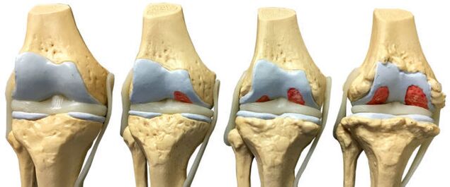
There are four stages of joint disease:
- The first stage is often overlooked. Sometimes morning stiffness and ankle pain may occur after strenuous exercise. When the feet move, a characteristic crunching sound will be heard. No pathological changes can be seen on the X-ray film, but the destruction process of cartilage has already begun.
- The morning stiffness was extended. The development of a leg takes 20-30 minutes. Limp sometimes occurs. Through the growth of bone tissue and the displacement of bones, the 2nd degree arthropathy of the ankle joint can be identified on the X-ray film.
- The symptoms of the three stages are obvious. Pain is no longer only after a heavy load, but also occurs during rest. Without painkillers, it is difficult for patients to do so. Limp increases. May need crutches. The affected joints are swollen and deformed. Ankle muscle atrophy. X-rays showed narrowing of the joint space, formation of osteophytes, and subluxation.
- The fourth stage is the most difficult. It developed due to lack of treatment. The cartilage is destroyed and the joint surface is fused. Walking is no longer possible.
With the development of ankle osteoarthritis, the cartilage and bone tissue of the articular surface gradually change.
diagnosis
The diagnosis of ankle joint disease is based on clinical symptoms and information obtained during the examination. Laboratory research is considered ineffective because there are no special tests that can detect pathology. In the remission period, all the indicators are within the normal range. As the condition gets worse, clinical blood tests will show high C-reactive protein and erythrocyte sedimentation rate. These indicators indicate that the pathological process has begun.
To confirm the diagnosis, use the instrument method:
- Radiography
- Magnetic resonance imaging;
- Ultrasound
- Bone imaging
- Diagnostic joint puncture.
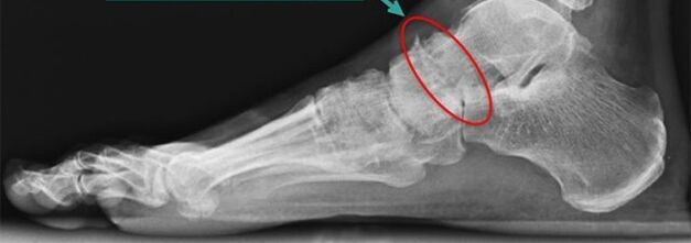
Simple radiography
Simple radiography is the most reliable and effective method for diagnosing diseases that occur in the musculoskeletal system. The principle of manipulation is the different absorption of X-rays by muscle tissue. Soft tissue allows X-rays to pass through, but hard tissue absorbs them. X-rays allow you to diagnose the disease itself and its consequences.
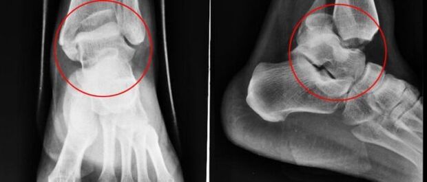
Conventional radiography is an inspection method in which a small amount of X-rays pass through the human body or part of the human body.
The snapshot allows you to view:
- The condition of the bone surface in the joint.
- The shape, size, and arrangement of the structures in the joint are interrelated.
- The condition of the fabric.
- The size of the joint space.
These indicators can help doctors determine the type and extent of joint damage. If the data is not enough, the doctor will prescribe other studies.
For ankle joint disease, X-rays will be performed in three projection methods:
- side;
- Back;
- Move the back foot inward.
The disease is characterized by the following changes:
- Reduce joint space;
- The presence of osteophytes;
- Osteochondrial replacement (subchondral sclerosis);
- Small spaces around the joints.
NMR
Nuclear Magnetic Resonance (NMR) is a diagnostic method that allows you to study the parts of your body that have water. The image shows bones in dark colors because they contain less water, but muscle tissue, intervertebral discs, and nerves appear lighter. MRI allows you to detect the smallest changes in bone tissue and joint structure. The study is also applicable to patients before joint prostheses. YMG has one disadvantage-high price.
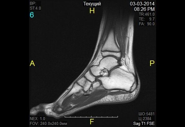
In nuclear magnetic resonance, the changes in the properties of hydrogen molecules under the influence of a strong magnetic field are recorded.
Magnetic resonance imaging
Magnetic resonance imaging (MRI) is an alternative diagnostic method that allows you to carefully examine the ligament structure of joints, muscles, and cartilage. With the help of MRI, the doctor assesses the condition of the calf joints. According to survey data, pathology is revealed in the early stages of development.
The diagnostic principle is based on exposure to radio waves and strong magnetic radiation. The magnetic field used is harmless and will not cause health hazards.
MRI is contraindicated in mental disorders, during pregnancy, and in the presence of metal objects in the human body.
When diagnosing ankle joint disease, use classic (closed) MRI machines because they have better image quality. The MRI machine is a large cylindrical tube with magnets around it. The patient is lying on a special table. The ankle is fixed with a special coil. This process takes 30-40 minutes. This study is absolutely painless. The patient may feel warm in the calf area.
Ultrasound
Since the 1990s, ultrasound has been widely used in medicine. This technique has proven its ability to make accurate diagnoses. Ultrasound scans are also performed for arthropathy of the ankle joint.
Today, ultrasonography is not particularly important in the diagnosis of osteoarthritis, because it does not allow adequate studies of damaged joints.
The equipment under study generates overclocking waves. The waves are reflected from the tissue and recorded on the monitor. Based on the obtained images, the doctor determines the type of pathology. In order to make the image on the monitor clear, a special gel is used. It eliminates the air gap and allows the sensor to slide better.
Ultrasound will not cause harm to the patient, so the process can be repeated many times. The advantages of ultrasound also include low cost and high precision.
The following indicators are obvious signs of joint disease:
- Cartilage thinning;
- The presence of bone growth;
- Joint effusion (synovitis);
- Loss of cartilage space.
Bone imaging
Scintigraphy is a high-precision study that uses isotopes to detect pathological changes in bones. The doctor divides the lesions into "cold" and "hot". In the first case, we are talking about regions without isotopes. Insufficient blood supply to these areas is not visible during the scan. The "cold" area is the place affected by malignant tumors. In the "hot" area, isotopes accumulate quickly, and they look very bright when scanned. These areas indicate the presence of an inflammatory process.
The role of scintigraphy in arthropathy is very important. When the clinical symptoms are extremely similar, the research helps distinguish arthropathy from many other diseases.
During bone scintigraphy, a special preparation containing specially labeled atoms is injected into the body.
Based on the results of the scintigraphy, the doctor makes a clinical prognosis and determines a treatment plan. The only drawback of this research is its high cost. Scintillation scanning is performed using special equipment, and unfortunately, not all medical institutions can afford it.
Although radiological scanning is a safe procedure, it still has many contraindications:
- pregnant;
- Lactation period
- Take barium-containing drugs.
When injecting radioactive substances, some patients experience allergic reactions such as itching and rashes. These side effects are not dangerous and will disappear on their own in a short time.
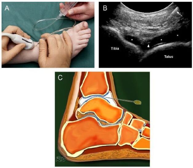
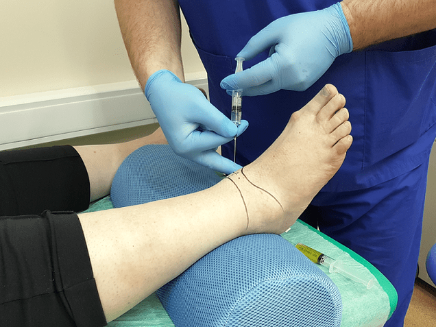
Joint puncture
Arthrocentesis is a diagnostic procedure in which a needle is inserted into the joint cavity to collect synovial fluid. This liquid was then sent for further study. Based on the data obtained, doctors make conclusions about the nature and stage of development of the disease.
At first glance, puncture is a simple procedure, but it is not. Extracting fluid from the joint capsule requires the doctor to act very accurately. The synovial membrane is very thin, and one awkward movement can cause trauma to it. As a result, an inflammatory process occurs. Potential risks also include infection. It is not difficult to bring infections into the joint capsule through poorly sterilized instruments.
The operation technique of each joint is different. When collecting joint exudate from the ankle joint, puncture the outer side of the ankle joint and the front between the extensor digitorum longus tendon.
Diagnostic sampling of intra-articular fluid allows laboratory analysis and rule out inflammatory arthritis.
Basic principles of treatment
After the diagnosis of ankle joint disease, the symptoms will not last long. Start treatment immediately. The further prognosis depends on the carefully selected treatment plan and the timeliness of initiation.
Arthropathy is an insidious disease. It cannot be cured completely. The goal of treatment is to stop the degenerative process and extend the remission period. To this end, doctors will prescribe medicine, physiotherapy, massage, gymnastics and folk remedies. If all the conditions are met, you can rely on positive motivation, otherwise the disease will progress.
Drug treatment of joint disease
According to the treatment effect, the drugs are divided into several groups:
- Anti-inflammatory drugs or painkillers. This group of drugs aims to eliminate inflammation and relieve pain. The earlier anti-inflammatory treatment is started, the greater the chance of salvaging the joint. This group of drugs can be made into tablets and ointments.
- Glucocorticoids. These drugs were prescribed when the aforementioned funds were invalid. They are produced in the form of a solution for injection. The medicine is injected directly into the joint.
- Cartilage protective agent. Designed to slow the destruction of cartilage.
The treatment plan and dosage are selected by the doctor according to the severity of the symptoms, the age of the patient, the presence of concomitant diseases and other factors. Self-medication is dangerous and often aggravates the condition, because many drugs have many side effects and have their own contraindications.

Features of radical treatment
If conservative treatment fails, then doctors will be forced to adopt radical treatment methods (surgical intervention). This action is also displayed in the following situations:
- Secondary (post-traumatic) and primary arthropathy of 3-4 degrees;
- Complications of arthropathy;
- Sustained severe pain in the ankle, radiating to the knee;
- Severe limp;
- Paralysis and paralysis of leg muscles;
- Violation of joint flexion and extension function;
- Violation of the supporting ability of the foot.
If the following conditions occur, surgical intervention is prohibited:
- The patient is under 12 years old;
- Fistulas are found in the joints;
- The patient has a history of diabetes and heart failure;
- An infectious disease was detected in the proposed intervention area.
Traditional treatment
Doctors believe that the treatment of joint disease should be completely under the supervision of specialists, but they do not deny the positive effects of folk remedies. Alternative medicine, as an effective preventive measure, helps to eliminate symptoms and maintain relief.

Folk remedies are symptomatic treatment of foot joint disease.
Home treatment should be coordinated with your doctor to avoid side effects and complications.
Traditional therapists recommend the following methods to treat the ankle joint:
- Burdock. Wash the burdock leaves with soap and running water. Apply the soft side of the leaf to the skin. Secure the top with a bandage or plastic wrap. It is best to keep it on all night.
- sea salt. Cut the salt in a saucepan. Pour it into a linen bag and tie it around your ankle. Hold the bag until the salt cools. Heat can relieve pain. Sand, lentils, and buckwheat are also used instead of salt.
- clove. Pour the triple cologne on the lilac flower. Leave the tincture in a cool place for 10-14 days. Rub the affected area sooner or later.
- eggshell. Grind the shells in a coffee grinder. Take out ½ teaspoon of the resulting powder. Before eating.
Don't forget that treatment with folk remedies should not be the only measure. Comprehensive treatment includes medication, exercise therapy, massage, physical therapy, and hydrotherapy. In advanced cases, doctors take radical measures-surgical intervention.
Surgery
For foot joint disease, the following types of surgery are used in medicine:
- Arthrodesis
- Arthroscopy;
- Endoprosthesis.
Arthrodesis is an operation that fixes joints. It is done to restore the limbs that have lost the ability to support. The only disadvantage of the operation is that the bones (tibia and talus) grow together, making it impossible to move. Arthrodesis is rarely used in medical practice.
Arthroscopy is a minimally invasive surgery. During the operation, the doctor makes small incisions in the joint area and inserts the arthroscopy (a special tube with a camera at the end) through these incisions. With its help, the surgeon carefully examines and evaluates the condition of the structures in the joints. If necessary, remove broken joints or blood clots from the synovial fluid. This operation is less traumatic. The only disadvantage of arthroscopy is that the risk of recurrence is too high.
The endoprosthesis is the last resort. It is performed in advanced arthropathy. The endoprosthesis allows you to partially or completely replace the affected joint. As a prosthetic product, innovative prostheses with modern mechanical structures are used. The service life of artificial joints is 10 to 20 years.
Power characteristics
In order to achieve good results, drug therapy is supplemented by diet therapy. Nutritionists have developed a special diet to prevent the disease from getting worse, while providing the body with all the necessary vitamins and nutrients. The diet of overweight patients plays a special role. Since obesity is one of the causes of joint disease, weight correction is an integral part of treatment.

The patient needs to reconsider some of his habits in daily life, these habits will lead to the development of foot joint disease.
Nutritionists recommend sticking to the following nutritional conditions:
- Eat often, in small portions.
- Drink at least 2 liters of fluids every day.
- Give up sweets and salt.
- The last meal is no later than 18. 00.
- Dishes can be steamed, boiled or grilled.
The main task of the joint disease diet is balanced and fortified nutrition. Fasting is impossible. A harsh diet and body cleansing do more harm than good. Calcium is excreted from the body, which is necessary for cartilage repair. A nutritionist will help you formulate your daily diet.
People with joint disease can eat grains, pasta, dairy products, cheese, beans, vegetables, fruits, rye bread, dried fruits, nuts, fish, and poultry. Heavy and fatty side dishes, foods containing dyes and spices, as well as kimchi, marinades, bacon, fatty broth, baked goods, spices, sauces, chocolate, ice cream, coffee, and alcohol are prohibited.
Prevent joint disease
To avoid the development of ankle joint disease, doctors recommend preventive measures:
- Wear comfortable heelless shoes;
- Keep on a diet and drink enough fluids;
- Take vitamin and mineral complexes seasonally;
- swim;
- Walk more in the fresh air;
- Eliminate excessive pressure on the legs;
- Avoid hypothermia;
- Ask a doctor to check in time.
For existing joint diseases, it is recommended to correct the lifestyle:
- Reject bad habits. They have been shown to cause stagnation of blood in the tissues and accelerate the destruction of cartilage.
- Perform a set of exercises to warm up your ankles.
forecast
Arthropathy is a progressive disease. If not treated, it will lead to irreversible consequences and complete immobility of the joints. Early diagnosis of pathology saves you from taking radical measures. Drugs can suspend the pathological process and relieve the patient's condition. There are no complications in the fight against the disease in the early stages.